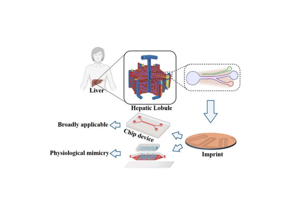
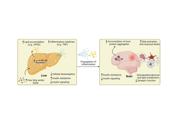
ORGAN MODELS

Liver

Liver-on-a-Chip constructs a three-dimensional microenvironment encapsulating hepatocytes and non-parenchymal cells (e.g., Kupffer cells, hepatic stellate cells) on a microchip platform, thereby recapitulating the physiological structure and functional characteristics of the human liver. This in vitro model provides a robust experimental tool for systematic investigations into liver function regulation, drug-induced toxicity evaluation, drug metabolic profiling, and the pathogenic mechanisms underlying liver diseases.

Cerebrovascular Nerve

The cerebrovascular-nerve organ-on-a-chip constructs a functional three-dimensional microenvironment by integrating multiple cell types (e.g., neurons, endothelial cells, pericytes, astrocytes) on a microchip, which can recapitulate the structural integrity and biological functions of the blood-brain barrier (BBB). Through the combination of microfluidic technology and biotechnology, this in vitro model effectively simulates the structural complexity of the human cerebrovascular system and its intrinsic neural functional characteristics, such as enabling the quantitative evaluation of BBB permeability and real-time monitoring of neuronal electrophysiological activities.It serves as a powerful tool for studying neurodegenerative diseases such as Alzheimer's disease, Parkinson's disease, and amyotrophic lateral sclerosis—facilitating investigations into pathological processes including neurofibrillary tangles formation, α-synuclein aggregation, BBB disruption-mediated neuroinflammation, and neuronal loss, as well as supporting preclinical evaluation of neuroprotective agents and targeted drug delivery systems across the BBB.

Cardiovascular System

By integrating microfluidic technology, biomaterial science, cell culture, and imaging techniques, the cardiovascular organ-on-a-chip constructs a biomimetic micro cardiovascular system with physiological structure and functional characteristics. This in vitro model recapitulates key dynamic physiological processes of the heart, including tissue-specific architecture, hemodynamic shear stress, transmural pressure, mechanical stretching, and electrical stimulation. It also enables the simulation of pathological states such as myocardial infarction and heart failure, while supporting quantitative detection of cardiac electrophysiological signals (e.g., action potentials, electrocardiographic-like waveforms) and analysis of electrophysiological remodeling. As a powerful experimental platform, the cardiovascular organ-on-a-chip provides an important tool for preclinical drug screening, toxicity evaluation, and cardiovascular disease mechanism research.

Reproductive Organs

The reproductive organ-on-a-chip is an advanced in vitro biomimetic model constructed by integrating microfluidic technology and MEMS (Micro-Electro-Mechanical Systems). Designed to recapitulate the complex physiological microenvironment and pathological progression of the human reproductive system, this platform regulates key culture parameters—including cytokine gradients, fluid shear stress, and hormone concentration dynamics—to faithfully reproduce the three-dimensional structural characteristics and tissue-specific dynamic functions of reproductive organs (e.g., ovaries, testes, endometrium). Beyond enabling mechanistic research on reproductive system diseases such as polycystic ovary syndrome, endometriosis, and male infertility, it also supports applications such as high-throughput screening of reproductive toxicants, evaluation of contraceptive drug efficacy, prediction of embryo implantation compatibility, and optimization of assisted reproductive technology (ART) protocols. As a cutting-edge experimental tool, it addresses the limitations of traditional in vitro models and provides novel insights for reproductive medicine research and translational applications.

Tumors

Tumor organ-on-a-chip models, tailored for breast cancer, lung cancer, colorectal cancer, and other malignancies, serve as powerful tools for investigating the initiation, progression, invasion, and metastasis mechanisms of tumors. Specifically, these biomimetic platforms enable the simulation of tumor-endothelial cell crosstalk to dissect the molecular and cellular mechanisms underlying tumor angiogenesis, while facilitating the real-time observation of tumor cell migration and invasive behavior within a physiologically relevant three-dimensional (3D) microenvironment. Beyond unraveling the pathogenesis of tumorigenesis, tumor organ-on-a-chip models exhibit broad applications in preclinical research, including high-throughput screening of novel antitumor agents, evaluation of drug efficacy and toxicity, and optimization of personalized tumor treatment regimens.

Skin

Skin-on-a-chip models typically integrate multiple cell populations—including keratinocytes, fibroblasts, and endothelial cells—to recapitulate the multi-layered anatomical structure and physiological functions of native human skin. In recent years, the integration of induced pluripotent stem cells (iPSCs) has significantly enhanced the physiological relevance of these biomimetic platforms, enabling them to closely mimic the phenotypic characteristics and functional properties of real human skin. Owing to their superior biomimetic performance, skin-on-a-chip models have been widely applied in evaluation of drug toxicity, quantitative analysis of transdermal drug penetration, efficacy and safety assessment of cosmetics, and modeling of skin-related diseases (e.g., atopic dermatitis, psoriasis, and skin cancer).

Rare Diseases

As an advanced microfluidic technology-driven platform, organ-on-a-chip enables the faithful simulation of the physiological microenvironment and pathological progression of human organs, thereby serving as a crucial tool for rare disease research. This technology exhibits considerable potential in spinal muscular atrophy (SMA) and Duchenne muscular dystrophy (DMD), by recapitulating disease-specific phenotypic traits and molecular pathways that are difficult to replicate with traditional models.

Liver-Brain Axis

On one hand, abnormal hepatic metabolism—such as ammonia accumulation and excessive release of inflammatory cytokines—can impair cerebral function, resulting in cognitive deficits, emotional disturbances, and other neurological manifestations. On the other hand, the central nervous system regulates hepatic metabolic processes (e.g., glucose and lipid metabolism) primarily through the vagus nerve, while chronic stress has been shown to exacerbate liver injury by disrupting this regulatory axis. As a biomimetic in vitro platform that recapitulates the bidirectional crosstalk of the liver-brain axis, the liver-brain axis organ-on-a-chip provides a powerful tool for identifying and validating drug targets associated with hepatic encephalopathy, Alzheimer's disease, and metabolic disorders linked to liver-brain dysfunction.

Gut-Brain Axis

Studies have demonstrated that the gut microbiota engages in bidirectional crosstalk with the central nervous system (CNS) via metabolites (e.g., short-chain fatty acids, bile acids), neurotransmitters (e.g., serotonin, γ-aminobutyric acid/GABA), and the intestinal barrier. This crosstalk modulates the functional states of CNS-resident glial cells, including microglia, astrocytes, and oligodendrocytes, thereby regulating neuroinflammatory responses and the progression of neurorelated diseases. The gut-brain axis organ-on-a-chip leverages microfluidic technology to recapitulate the multi-component interactions among gut microbiota, intestinal epithelial barrier, blood-brain barrier (BBB), and neural tissue in a biomimetic 3D microenvironment. This advanced in vitro platform provides a unique tool for dissecting the underlying mechanisms of gut microbiota-mediated neurodegenerative diseases (e.g., Alzheimer's disease, Parkinson's disease) and supports the discovery and preclinical evaluation of novel therapeutic agents targeting the gut-brain axis.

Heart-Brain Axis

The heart-brain axis denotes the intricate network of connections between the heart and the brain, mediated through neural, humoral, immune, and other regulatory pathways. Chronic hypertension elevates the risk of stroke, while stroke can conversely induce cardiac dysfunction; cardiac diseases may also contribute to cognitive decline via mechanisms such as impaired cerebral blood perfusion. The heart-brain organ-on-a-chip model enables systematic investigation of bidirectional heart-brain crosstalk, including the effects of myocardial inflammation on neural function, stroke-induced cardiomyocyte apoptosis, and the reciprocal interactions between neurodegenerative diseases (e.g., Alzheimer's disease) and cardiac disorders. By recapitulating the physiological and pathological interplay of the heart-brain axis in a biomimetic in vitro setting, this platform provides a powerful tool for dissecting underlying disease mechanisms and supporting preclinical development of targeted therapies for heart-brain comorbidities.

Heart-Brain-Liver-Kidney

The heart-brain-liver-kidney multi-organ-on-a-chip exhibits substantial application value in preclinical drug toxicity prediction (particularly cardio-cerebral neurotoxicity), complex disease mechanism dissection, and personalized medicine advancement. Specifically, the integrated hepatic module recapitulates drug metabolic processes (e.g., phase I/II biotransformation), while the renal module simulates drug excretion pathways (such as glomerular filtration and tubular secretion). Meanwhile, the cardiac and cerebral modules enable quantitative evaluation of drug-induced effects on cardiac function (e.g., arrhythmia, QT interval prolongation) and nervous system integrity (e.g., neurotoxicity, synaptic dysfunction). By recapitulating inter-organ crosstalk in a biomimetic microenvironment, this platform facilitates real-time and dynamic investigation of the synergistic or adverse effects of drugs across multiple core organs, addressing the limitations of single-organ models and providing more clinically relevant data for drug development.